Introduction
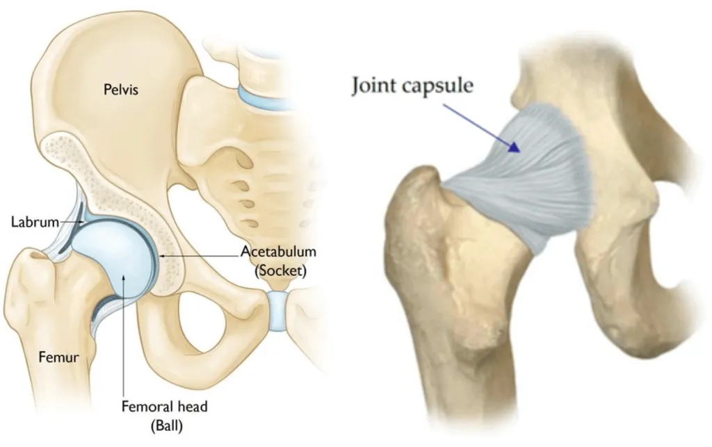
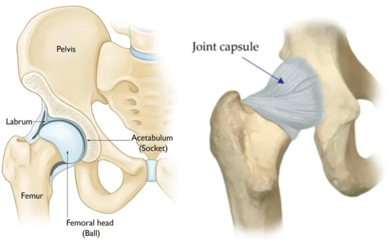
The hip joint is one of the most important and largest joints in the human body. It is a ball-and-socket joint that allows a wide range of movement. The hip joint consists of two main parts: the ball (femoral head) at the top of your thigh bone (femur), and the socket (acetabulum) in your pelvis. These parts work together to enable movements like walking, running, and jumping.
In this article, we will break down the anatomy and key functions of the hip to help you understand more of how it works!
Bones of the hip
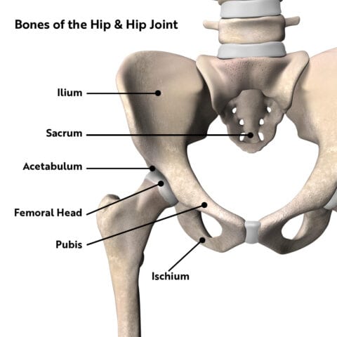
The hip is a ball-and-socket joint that involves three primary bones. These include the femur (the thigh bone), the pelvis, and the hip bone itself. The pelvis is a complex structure made up of several parts, including the ilium, ischium, and pubis. The femur’s head fits into a socket in the hip bone, known as the acetabulum, forming the hip joint. This joint is stabilised by ligaments and moved by various muscles, allowing for a wide range of motion.
Ligaments and capsules of the hip
The ball-and-socket joint is surrounded by a complex network of ligaments and capsules to provide stability. These structures prevent the hip from dislocating, while also allowing a wide range of motion.
The ligaments of the hip are tough, fibrous tissues that connect the bones of the hip joint. There are three main ligaments in the hip:
Iliofemoral ligament is the strongest ligament in the body. It connects the pelvis to the femur (thigh bone) and prevents excessive extension of the hip.
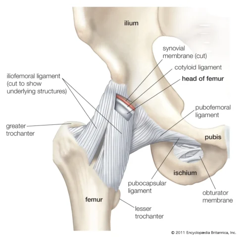
Pubofemoral ligament also connects the pelvis to the femur, but it primarily restricts excessive abduction and rotation of the hip.
Ischiofemoral ligament restricts internal rotation and extension of the hip.
The hip joint is also surrounded by a capsule, which is a thick, fibrous structure that encloses the joint. The capsule is lined with a thin layer of synovial tissue that produces synovial fluid. This fluid lubricates the joint, reducing friction and allowing smooth movement. The ligaments and capsule of the hip work together to maintain the stability of the joint, while also allowing the extensive movement needed for activities like walking, running, and jumping.
Muscles and tendons of the knee
Muscles are tissues that contract to cause movement, while tendons are strong, flexible bands of tissue that connect muscles to bones.
Connecting these muscles to the hip bone are several tendons. Tendons attach muscles to the bones. They distribute the force from the muscles through our body evenly by absorbing the impact and propelling it back through the ground as we walk or run.
There are several key muscles in the hip area that provide function in various ways such as:
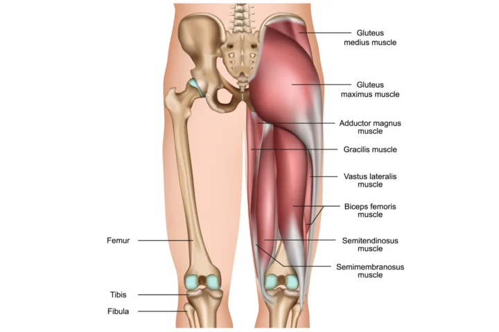
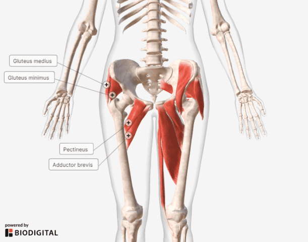
The primary extensors are Glute Max. The secondary extensors include the hamstring muscles (Biceps Femoris, Semitendinosus and Semimembranosus). They are important propelling us forward (for walking and running) and up (squatting, jumping and hopping).
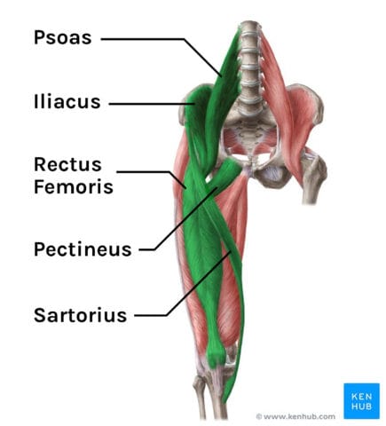
The primary flexors are Rectus femoris, Iliacus, Psoas, Iliocapsularis, and Sartorius. The secondary flexors are Psoas minor and Pectineus. They are important for walking, lifting the knee and pelvic stabilisation.
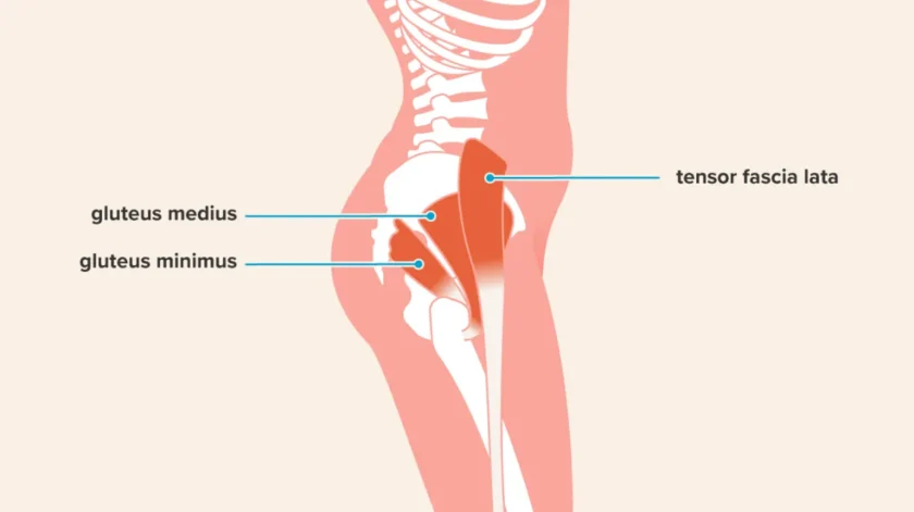
The primary abductors are gluteus medius, gluteus minimus and Tensor Fasciae Latae (TFL). The secondary abductors include piriformis, sartorius and superior fibres of the gluteus maximus. They are important for walking and pelvic stabilisation.
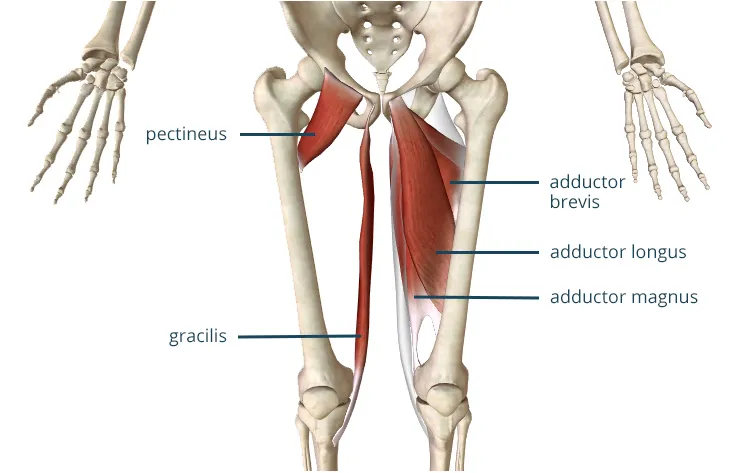
The adductors consist of 5 muscles which are divided into long and short adductors. The long adductors are Gracilis and Adductor Magnus which attach to the pelvis extending to the knee. The short adductors are Pectineus, Adductor Brevis and Longus which attach at the pelvis and extend to the thigh bone.
The primary hip internal rotators are TFL, Glute Medius and Minimus, Adductors (groin). The secondary internal rotators include Glute Medius and Mininimus, TFL, Adductors (groin). They are important for gait efficiency and hip mobility through wide range of movements in the gym via pelvic stabilisation.
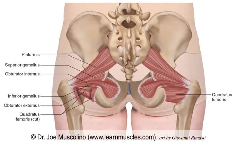
The primary hip external rotators is the Piriformis, Glute Max. The secondary external rotators include Super Gemellus, Obturator Internus, Inferirior Gemellus, Obturator Externus, Quadratus Femorus. They are important for maintaining hip, knee and ankle alignment alongside generating torque at the hip joint for performance benefits.
Functions of the knee
The function of the hip is to support your body when you stand and move, making sure you’re stabilised and balanced. The hip allows us to perform a wide range of movements whether they are short and stable or powerful large movements. Ensuring appropriate strength and flexibility (isolated) and global mobility, stability and strength will allow you to perform optimally whilst reducing the risk of injury in your chosen sport, physical activity or exercise method.

Summary
The hip joint plays a crucial role in supporting the weight of the body in both static (standing) and dynamic (walking or running) postures. This is done so by a variety of movements including: flexing, extending and rotating the leg, transferring force from lower to upper body and vice versa.
Stay tuned for our next article to find out how to strengthen these structures and how to reduce your future injury risk.
