Introduction
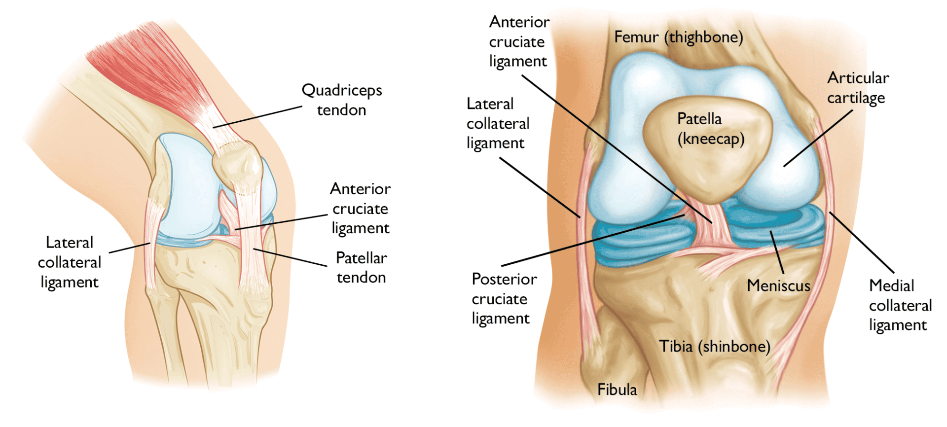
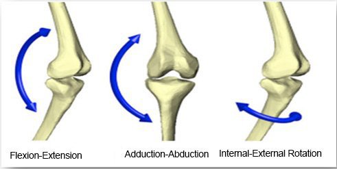
The knee is joined together at the tibia and femur and the femur and patella. It is the largest joint in the human body and is also a synovial hinge joint which means that it allows you to move freely in one direction (up and down) with a slight rotation of the tibia on end-range flexion and extension. The knee allows you to bear the force of the ground to your upper body and helps move and bend your legs.
In this article, we will break down the anatomy and key functions of the knee to help you understand more about how you stand.
Bones of the knee
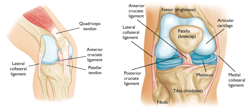
The knee is joined by 3 bones, tibia (shin bone), femur (thigh bone) and patella (knee cap). It is also known as the tibiofemoral joint. There are 2 articulations (where the bones meet) in your knee, Patellofemoral is when the patella meets the femur and tibiofemoral is when the femur meets the tibia.
The femur and thigh bone connect the hip to the knee, the tibia connects the knee to the ankle. The patella (kneecap) is a small bone in front of the knee and sits on the knee joint as the knee bends allowing the movement to run smoothly.
Ligaments and capsules of the knee
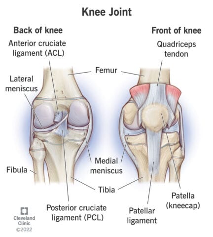
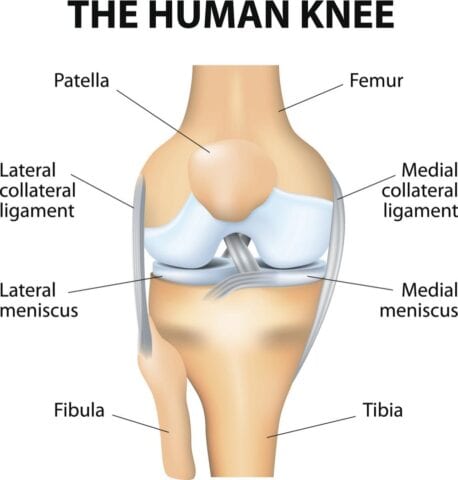
The cartilage acts as a shock absorber for the knee, there are 2 types; hyaline cartilage which covers the top and bottom of the bones to make sure they glide smoothly across one another and fibrocartilage which is a tough cartilage holds parts of the body in place and absorbs impact. The meniscus is 2 parts of the fibrocartilage and cushions the space between the femur and tibia.
The knee contains 2 types of ligaments: Collateral ligaments which are your lateral collateral ligament (LCL) and medial collateral ligament (MCL). They are situated on the outside of your knee and connect your femur to your calf bone (fibula) preventing your knees from over-extending side to side.
Cruciate ligaments which consist of 2 of them in your body. They cross over at the front and the back which prevents the knee from moving too much forwards and backwards. The anterior cruciate ligament (ACL) crosses at the front and the posterior cruciate ligament (PCL) crosses at the back.
Muscles and tendons of the knee
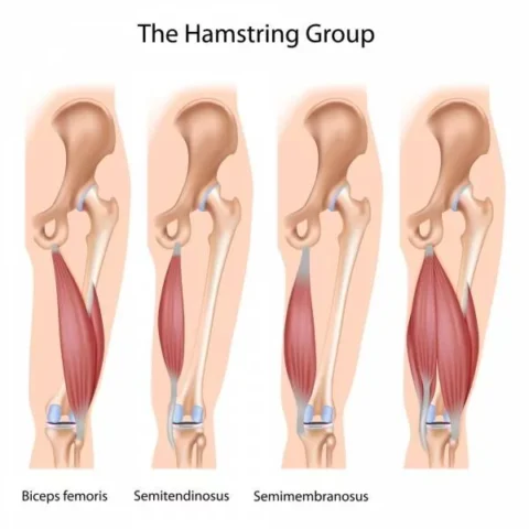
The muscles in the knee are important in stabilising the joints for movement. Each muscle has its function and supports the knee. The hamstring muscles are situated at the back of the thigh that helps the hip extend (go behind you and the knee to bend. They start from the bottom of the pelvis (ischium) and attach behind the knee (at the tibia). They are made up of 3 muscles; Bicep femoris (the long muscle that flexes the knee), semimembranosus (is also a long muscle that extends the thigh, flexes the knee and rotates the tibia. Lastly, Semitendinosus (also extends the thigh and flexes the knee but has narrower tendons unlike semimembranosus).
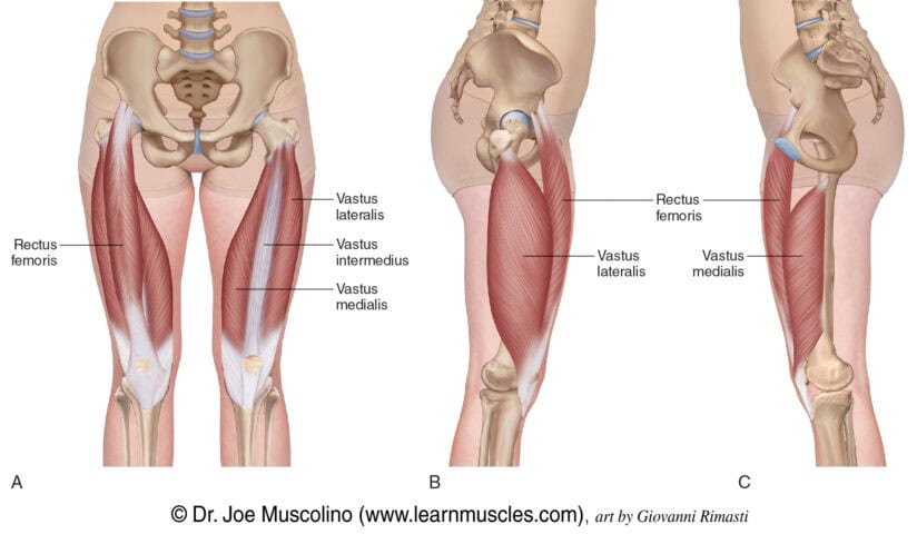
The quadricep muscles are made up of 4 muscles which are; rectus femoris, Vastus Lateralis, Vastus Medialis and Vastus Intermedius. The rectus femoris attaches to the knee cap (patella) and has the least effect on extending the knee but assists in hip flexion. Vastus Lateralis is on the outside of the thigh and attaches at the top of the femur at the hip and to the knee cap. It is also the biggest out of the muscle group, Vastus Medialis aids knee extension and is also known as the tear-shaped muscle which sits at the inner thigh and attaches along the femur and down to the inner border of the knee cap. The Vastus Intermedius is the deepest muscle and is situated at the front and between Vastus Lateralis and Vastus Medialis.
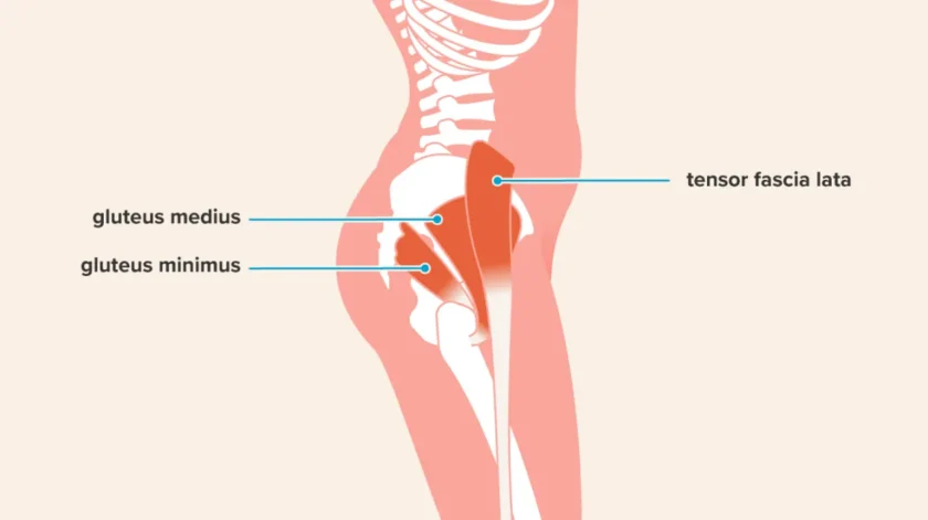
The glutes consist of the minimus, maximus and medius. The gluteus maximus is attached to the lower body by the iliotibial band (IT band) which is located at the side of the thigh. If the glutes are weak then the thighs have a higher risk of rotating inwards.
The primary abductors are gluteus medius, gluteus minimus and Tensor Fasciae Latae (TFL). The secondary abductors include piriformis, sartorius and superior fibres of the gluteus maximus. They are important for walking and pelvic stabilisation.
The adductors consist of 5 muscles which are divided into long and short adductors. The long adductors are Gracilis and Adductor Magnus which attach to the pelvis extending to the knee. The short adductors are Pectineus, Adductor Brevis and Longus which attach at the pelvis and extend to the thigh bone.
Functions of the knee
The function of the knee is to support your body when you stand and move, making sure you’re stabilised and balanced. The knee allows us to perform a wide range of movements whether they are short and stable or powerful large movements. Ensuring appropriate strength and flexibility (isolated) and global mobility, stability and strength will allow you to perform optimally whilst reducing the risk of injury in your chosen sport, physical activity or exercise method.
Summary
The knee is a very complex joint and it is a huge factor in our everyday lives and physical activity, providing us with support, stability, strength and force distribution.
Stay tuned for our next article to find out how to strengthen these structures and how to reduce your future injury risk!
