Introduction
The ankle joint is made from the lower leg and foot and is a key requirement for gait and other activities of daily life. It is a hinged synovial joint that joins the talus, tibia and fibula bones. The ankle plays a key role in maintaining stability while transporting force from the ground up the body and back into the ground.
In this article, we will break down the anatomy and key functions of the lower limb to help you understand more of what makes you stand tall!

Bones of the ankle and the foot
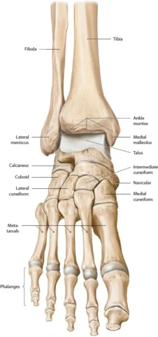
The ankle is joined by 3 bones, the talus, tibia and fibula. The complexity of the ankle is formed by 3 sub joints: talocrural, distal tibiofibular and subtalar joints. The stronger and larger bone out of those 3 is the tibia, which forms the inside part of the ankle (shin bone). The fibula is the smaller bone of the lower leg that forms the outside part of the ankle. The talus is the small bone between the tibia and fibula and the calcaneus is also known as the heel bone.
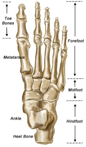
The foot can also be divided into 3 parts; the hindfoot, the midfoot, and the forefoot. The hindfoot is composed of 2 of the 7 tarsal bones. The talus and the calcaneus are the midfoot containing the tarsal bones and the forefoot contains the metatarsals and the toes (phalanges).
Ligaments and capsules
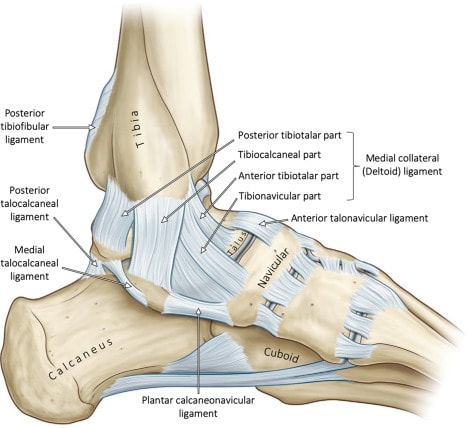
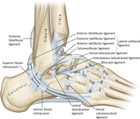
Many ligaments hold the ankle joint together (this is to maintain the joint stays intact and prevents it from coming apart). The lateral (outside) ligaments in the ankle include the anterior talofibular ligament (ATFL), Calcaneofibular ligament (CFL) and posterior talofibular ligament (PTFL). ATFL connects the front of the talus bone to the fibula. The CFL connects the calcaneus to the fibula and the PTFL connects the rear of the talus bone to the fibula. As a unit, these ligaments provide support to the outer ankle and are often the main injured tissues when we roll or sprain an ankle.
There is also a thick ligament called the deltoid ligament which supports the entire medial/inner side of the ankle. This is made up of the anterior tibiotalar ligament (ATTL), posterior tibiotalar ligament (PTTL), tibiocalcaneal ligament (TCL) and tibionavicular ligament (TNL). The anterior (front) inferior (lower) tibiofibular ligament (AITFL) connects the tibia to the fibula.
Two ligaments that crisscross the back of the tibia and fibula are the posterior inferior tibiofibular ligament (PITFL) and the transverse ligament. The interosseous ligament rests between the tibia and fibula and runs the entire length of the tibia and fibula, from the ankle to the knee.
The multiple ligaments that surround the ankle form part of the joint capsule. This is a fluid-filled sac that surrounds and lubricates the joints.
Muscles and tendons of the ankle
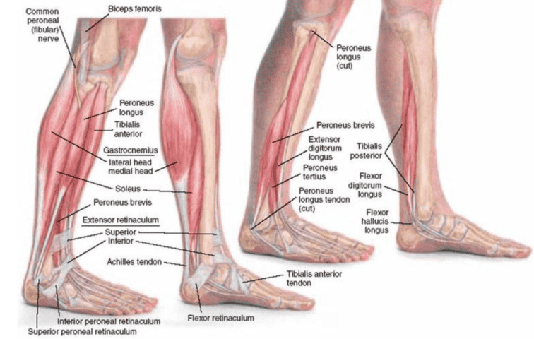
The main muscles in the ankle are; the Gastrocnemius and soleus which support plantar flexion (pointing the foot away) with support of the posterior tibialis which supports the foot’s arch. The anterior tibialis allows the foot to move upwards (dorsiflexion) and inversion (with the assistance of the hallucis longus muscle). Peroneal tibialis controls the movement on the outside of the ankle (eversion) with peroneus tertius. Peroneal tibialis controls the movement on the inside of the ankle (inversion) with peroneus tertius.
The main tendons are; the Achilles tendon which attaches the calf muscles (gastrocnemius and soleus) to the heel bone allowing you to perform movements such as walking, running, and jumping. The posterior tibial tendon attaches one of the smaller muscles of the calf to the underside of the foot and helps support the arch allowing us to turn the foot inward. The anterior tibial tendon allows us to raise the foot which is done by two tendons that run behind the outer ankle and help us turn the foot out (eversion).
Functions of the ankle
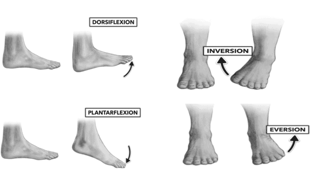
The function of the ankle is to perform movements in six different ways; dorsiflexion, plantar flexion, inversion, eversion and medial and lateral rotation. By doing those movements it supports the body and maintaining balance. The final key functions to note are the ability to transfer force by propulsion (allowing us to propel the body forward) when walking or running by pushing off the ground and shock absorption which protects the joints and bones in the foot and leg from impact.
Summary
As you can see, the ankle and foot is made up of many muscles, ligaments and tendons to protect the bones on impact and helps with everyday activities and exercise due to the stability it provides. Stay tuned for our next article to find out how to strengthen these structures and how to reduce your future injury risk.
